

It is just over a century since Perthes disease (also known as coxa plana, Legg-Calvé-Perthes, Legg-Perthes, and/or Legg-Calvé), a disease that affects the hips in children, was first identified. It remains extremely rare, and indeed it is growing rarer. Many practitioners do not register that this may be why children are in considerable pain and/or have difficulty walking, sometimes for years. Indeed, even when children are diagnosed, they may receive any one of a whole number of treatments.
The patient profile
The disease pattern is still not fully understood; but, very broadly, there seem to be environmental rather than genetic factors involved. It mainly affects boys (around 80% of cases). Worldwide, studies to date show that the greatest incidence is in northern Europe, with the incidence rising sharply by around nearly 50% with every 10 degrees further north from the equator. In the UK, it is much more prevalent in the northern regions.
There also appears to be a familial but not a genetic element involved: research on twins showed that if one twin has the disease, the chance of the other twin being affected is no higher in identical twins compared to non-identical twins, but there is a slightly higher likelihood that a family member will be affected compared to the normal population (Metcalfe et al, 2016). There is also a link with social deprivation, with deprived children being much more likely to develop it (Perry et al, 2012).
‘Many practitioners do not register that [Perthes disease] may be why children are in considerable pain and/or have difficulty walking, sometimes for years. Indeed, even when children are diagnosed, they may receive any one of a whole number of treatments’
Children usually first present with pain in their hips, groin, legs and/or knees (this may be referred hip pain). They may also be limping, and/or find it difficult to get the full range of movement with their hips. It may come on suddenly or have been happening for some time.
They also have other characteristics. They are usually shorter than average, and their growth has usually slowed further in the year before they present. They tend to be more active than other children and they may also have a genitourinary abnormality such as a hernia, undeveloped testes or hypospadias.
Bone development
Perthes affects the blood supply to the hip joint: specifically, to the rounded head of the femur where it fits into the hip socket, and to the growth plate (physis). Over time, this blood loss means that the bone starts to soften and break until the blood vessels regrow and new bone is produced. This may take several years – and importantly, the new bone growth may not be in the ‘right’ shape to fit properly into the hip socket.
The bone changes go through the initial necrosis, sclerosis, fragmentation and reossification phases, before reaching the final ‘healed’ stage. At the very beginning, even though the bone is softening and causing pain, it is quite hard to detect under an X-ray and can only be identified using a magnetic resonance imaging scan. As the sclerosis progresses, the dead bone becomes detectably white under an X-ray, and starts to flatten. After that, as the new blood vessels form, the dead bone is broken down and reabsorbed while new bone is formed from the outside in. In the final reossification stage, the central bone of the new femoral head is formed.
There are two main problems. One is the pain and mobility difficulties that the young patients usually experience while the hip is unable to work properly. The other is that the new bone does not always reform into the original rounded shape. It may be a more oval ‘rugby ball’ shape or quite flat. The more it diverges from a ball shape, the less likely it is to fit properly into the hip socket, affecting mobility permanently and also causing pain. A significant proportion of adults who had Perthes as children need a hip replacement in later life.
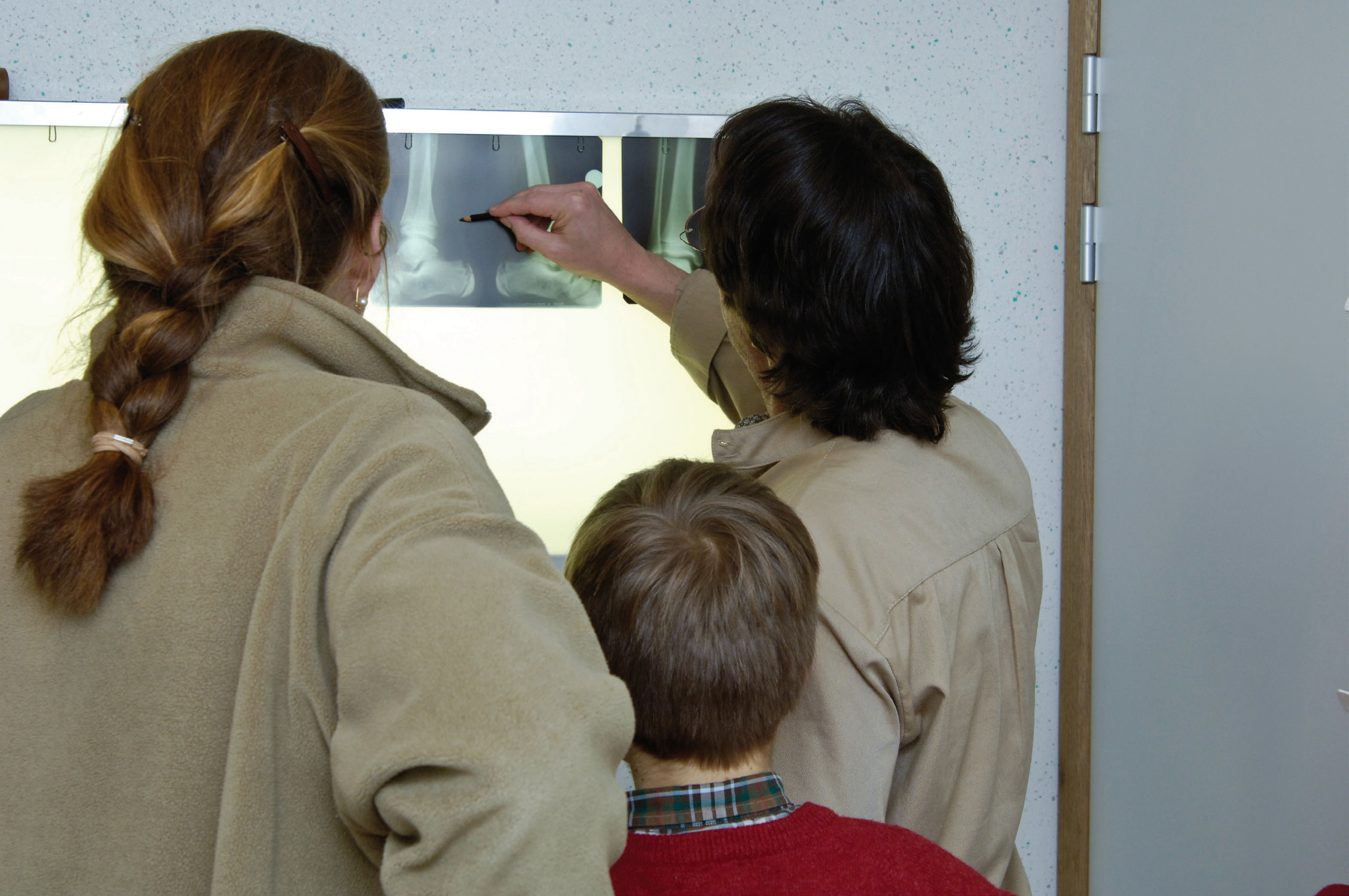 Perthes disease affects children's hips and causes pain and mobility difficulties. It is very rare and usually diagnosed by X-ray. Treatment for Perthes disease usually focuses on pain management, and can range from a ‘wait and see’ approach, to surgery on the hip and pelvis.
‘Perthes is usually diagnosed by X-ray (occasionally magnetic resonance imaging is also needed). The treatments focus both on pain management and on gett ing the bone to reform properly.’
Diagnosis and treatments
Perthes is usually diagnosed by X-ray (occasionally magnetic resonance imaging is also needed). The treatments focus both on pain management and on getting the bone to reform properly. Most children are under some kind of pain management regime; but it is important to flag up with parents, carers and/or other professionals if they seem to be experiencing a lot of pain despite this.
Th ere is a range of around 10 treatments, from a ‘wait and see’ conservative approach, through casts and braces, to different forms of surgery on the hip or pelvis to adjust the positioning and ensure that the hip joint functions as easily as possible. The stages to concentrate on are the early ones, up to the early stages of fragmentation, when the bone is still quite soft and can still be influenced. But the choice of treatment really depends on the unit where the child is being treated. ‘Some treatments are almost the opposite of another and surgeons may hold completely different beliefs about how a patient should be treated. In practice, we tend to go with the way that historically our unit has practised,’ explained Daniel Perry (personal communications, 2021), during a phone interview in March. Daniel Perry is the Associate Professor of Orthopaedics and Trauma Surgery at Oxford University's Nuffield Department of Orthopaedics, Rheumatology and Musculoskeletal Science, as well as an honorary consultant Children's Orthopaedic Surgeon at Alder Hey Hospital.
Management
Some children are admitted to hospital for bed rest and/or traction using light weights. However, it is necessary to keep children as active as possible, in order to maintain a good degree of strength and muscle length in the hip joint. This is particularly important to avoid pulling the hip into a position where, with the increased load, the hip joint starts to collapse even further. This means encouraging the most movement possible – football is still an option – but activities that involve joint force should be avoided.
Surgery
Surgical treatment is less common in Europe than North America; the most common operation is femoral osteotomy, but pelvic osteotomy is comparably more common in North and South America and in Australia (Braito et al, 2021). Yet the outcomes appear to be fairly similar. Overall, surgery is marginally more likely to result in a well-formed bone shape. The ‘Herring study’, a cohort study conducted in 2004 looking at 438 patients with 451 affected hips, is the main study to date on the effectiveness of different approaches (Herring et al, 2004). It found that surgery had a 60% chance of the best (rounded) hip shape and 10% of the worst (flattened), while no surgery was associated with 45% best and 20% worst. Around a third in both cases – 30% with surgery, 35% without – were in the middle, with a shape that was neither round nor flat.
‘But these differences aren't huge,’ Perry points out. ‘Surgery isn't a miracle cure; the differences are marginal at best. It carries some level of risk and usually involves two operations – one to do the operation, and one later to take the metal plates, screws or wires out. Children usually wear casts for a period afterwards. There's quite a big insult to the child, but there's also a duty on the part of the surgeons to do everything you can for that child. Often children undergo surgery primarily because we're worried about not doing enough if we don't’ (personal communications, 2021).
Research in progress
This is a condition about which there has been relatively little research to date, but this is slowly changing. The James Lind priority-setting partnership of surgeons and families has made research into Perthes a priority (James Lind Alliance, no date), as has the British Society for Children's Orthopaedic Surgery (Perry et al, 2018).
Perry is designing a randomised control trial, to build on the Herring study which is, as he says, ‘the best we can hang our hats on’ at the moment. This is quite a new move in surgery, but randomised controlled trials have been getting under way in areas like fractures, and surgical teams are now coming on board with conducting one for Perthes. ‘All the work I've done beforehand combining different trials and research has been pointing towards the ultimate randomised trial in Perthes disease’ (Perry et al, 2018).
Adam Galloway is a paediatric physiotherapist and Visiting Academic Clinical Fellow at the University of Leeds. He has just secured funding from the National Institute for Health research to complete a PhD on developing a new complex intervention in the form of an app. In an interview in March, he explained that his research is split into three workstreams (personal communications, 2021). ‘One, speaking to key stakeholders (children and family's clinicians, clinical nurse specialists, physios, surgeons and so on) to discuss the current experiences of non-surgical care and doing qualitative interviews and identify gaps in non-surgical treatment. In the second, I'll be working with NHS Digital, and a local app developer to develop the app. We don't know what that will look like yet, but it should make it possible both for people to access information about the disease and for children to log their exercise and other progress. Finally, we'll be testing it with a small group and patients and families, to assess how useful it is as a non-surgical treatment’.
‘Historically, Perthes disease is supposed to cause a “painless limp’’,’ Perry concluded in his interview. ‘We know that's not true. There's not enough known about the disease at primary care level and there is too much variation at secondary care. We need to change that, because the variation is unacceptable.’
FURTHER INFORMATION
Steps
For more information visit: www.stepsworldwide.org
Email: info@steps-charity.org.uk
Phone: 01925 750271